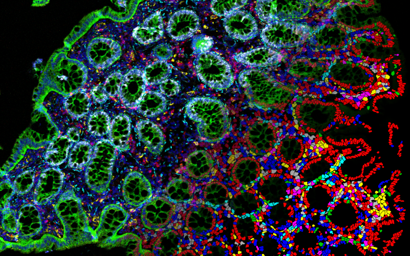
To understand how to best treat cancer, it is essential to study how cancer cells behave and organize. The relatively new field of ‘Spatial Omics’ provides valuable new insights by visualizing cancer cells in a unique way.
With spatial omics, various techniques are combined to study the position of cancer cells in tissue from cancer patients. Researchers sometimes refer to it as ‘spatial biology’. This helps to understand the origins of cancer and devise new treatments.
Associate professor Yvonne Vercoulen is a group leader at UMC Utrecht, and her team specializes in spatial omics. “My team develops new techniques that allow us to simultaneously view all kinds of different cells.” Yvonne and her team explain the latest developments.
Why Spatial Omics?
Cells can be seen as the LEGO bricks of our body. The organization and cooperation of our cells determine whether our body remains healthy. Yvonne compares it to a car made of LEGO bricks: “The LEGO bricks together create a functioning car. If the organization of the bricks is disrupted, the car falls apart or flies off the road. The same happens in your body: if the organization of cells is disrupted, damage occurs, and you can get sick.”
To understand how this works, it is important to accurately map the organization of our cells. This is where spatial omics comes in. Yvonne’s research group focuses primarily on the organization of immune cells, the cells that make up our immune system. This gives them new insights into cancer and immunological diseases, which are closely related. “These two fields are very much intertwined,” Yvonne explains. “For example, you see that people with too much inflammation have a higher risk of cancer. Conversely, tumor cells in turn affect our immune system to evade it.”
Distance Matters
Spatial omics is used, among other things, to predict which patients’ tumors will metastasize. Why is this important? “To prevent that patients receive unnecessary chemotherapy with all the associated side effects,” Yvonne explains. One of the projects focuses on a type of colon cancer that only metastasizes in 1 out of 5 people. For these metastases, these patients really need chemotherapy, but it is difficult to predict which patient will develop these metastases. “We looked into how immune cells organize around the tumor,” Yvonne explains. And what turns out? If immune cells are further away from the tumor and trapped in connective tissue, the risk of metastasis is greatest. “With this knowledge, patients can receive more targeted treatment, and overtreatment can be prevented.”
“We hope that spatial omics will help to choose the best therapy for patients and to devise new treatments.”
Quality of life for patients
Spatial omics is also used to predict which patients will benefit from certain therapies. Stephanie van Dam is a physician-researcher in Yvonne’s research group and focuses on a type of skin cancer, metastatic melanoma. These patients are treated with immunotherapy. This works very well in half of the patients, but in the other half virtually no patient benefits. Little is known about who will and will not respond to the therapy and the biology behind this. Stephanie uses spatial omics to gain more insight into this.
Stephanie: “For these patients, immunotherapy is their last chance, and it currently takes about three months to see if the therapy is working. That’s very long for these patients, who often don’t have a long survival time left.” Stephanie collects tissue from patients before they receive treatment. She then uses spatial omics to compare the position and the amount of different immune cells in this tissue. Based on this, she tries to predict which patient will of will not respond to the treatment. “This way, you save patients three months of unnecessary treatment and all the side effects that come with it.”
“I try to predict which patients will benefit from immunotherapy to avoid unnecessary treatment with all the side effects that come with it.”
Collaboration as a strength
Spatial omics requires the combination of many different techniques. For this, good cooperation is essential. Therefore, the Spatial Omics Accelerator was recently established, a collaboration between researchers in Utrecht. Kristof van Avondt (Assistant Professor) leads this new project. “Everyone has their own specialization. Through new collaborations, we can combine different techniques,” explains Kristof. “This allows us to build knowledge that various researchers can use, along with our techniques and advice, to answer their research questions.” Additionally, the Spatial Omics Accelerator offers opportunities for new innovations, providing even more insights into tissues through spatial omics.
“The amount of collaboration makes this work incredibly fun.” says Yvonne. “Also, the doctors here at UMC Utrecht think along about what is important for the patient.”
“With the new Spatial Omics Accelerator, we are constantly innovating and trying to contribute to the spatial omics of tomorrow.”
Where do you hope to be in five years’ time?
Yvonne: “I hope that our techniques will have contributed to improvements in the clinic. Hopefully, we can by then make better choices for the right therapies for patients and provide insights for new therapies.” Yvonne and her team also hope that their spatial omics techniques will be widely used in the research field.
What is spatial omics?
Spatial omics is essentially the spatial analysis of tissue by using a combination of different techniques. Thanks to many technological developments, we can now measure biological molecules in cells with a very high resolution. By measuring DNA or proteins in cells, we gain a lot of information about what types of cells they are and what state they are in. However, these techniques don’t tell us much about the spatial organization of the cells. Combining these techniques with imaging techniques gives information about the characteristics of the cells ánd their specific locations in the tissue. This results in beautiful images, but even more important it provides important insights into the biology of cancer. Yvonne: “We really need this unique combination of information to offer effective therapy to every patient.”
Image at the top is made by Matthijs Baars.


