AI helps to find breast cancer metastases faster and in a less expensive way
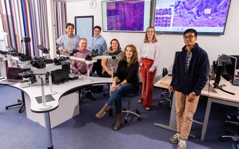
AI and pathologist team up
In healthcare, AI increasingly seems to be contributing to more efficient healthcare. At UMC Utrecht, we apply and research AI in multiple ways. Our pathologists, for instance, already use AI in daily standard practice to find metastases in case of breast cancer, helping them to work faster, more efficient and in a less expensive way.
After a woman’s breast tumor is removed for breast cancer, often the sentinel lymph node is also taken away. By doing so, the pathologist can check if the cancer has metastasized. That is, if breast cancer starts spreading, the tumor cells end up in that sentinel gland first, via the lymph fluid. That is why pathologists always carefully examine that tissue as well.
With about two-thirds of the patients, the pathologists have good news, because they have found no metastases in the sentinel gland. But to be really sure about this, pathologists take another look at sections (thin slices) of the glandular tissue. This time, they use additional staining, containing antibodies that recognize tumor-specific proteins.
“Because these antibodies have a color label, the tumor cells become clearly visible,” explains assistant professor Carmen van Dooijeweert.
“This way, you can recognize possible tumor cells, which might have been missed during the first assessment, without the extra staining. Those extra stains are quite expensive, about 25 euros per tissue section, of which we already do (at least) five per sentinel node. Also, sometimes multiple tissue blocks from numerous sentinel lymph nodes need to be viewed, and then the costs will rise even more rapidly. Furthermore, it is an intensive, time-consuming job for the pathologist.”
“Healthcare innovations are often expensive, but this innovation actually reduces healthcare costs. And AI allows pathologists to get on with their other work faster.”
Faster and less expensive
During the CONFIDENT-B trial, Carmen has investigated whether AI can help to perform sentinel node tissue assessment more efficiently. Can it be done faster and – with rising healthcare costs in mind – less expensive, without risking missing tumor cells when the extra staining is omitted?
“AI can be used in two ways: completely independent (‘independent AI’ – ed.) or as an assistant to the healthcare professional (‘AI assistance’ – ed.),” says Carmen. “With Independent AI, the algorithm decides independently whether something is a tumor or not. This is quite risky, because what if the algorithm is wrong? That raises all kinds of ethical and legal questions?”
Carmen and her team therefore use the other form of artificial intelligence, AI assistance: the algorithm lends a helping hand, but you will still have to take a look at the result yourself.
“Our algorithm – which we did not develop ourselves but we licensed it from the company Visiopharm – recognizes structures,” Carmen says. “It pinpoints anything which doesn’t belong in a lymph node and marks it for the pathologist. The algorithm then puts a red, orange or yellow outline around it, depending on how suspicious it thinks the spot is.”
Those markings are a signal to pathologists that they will need to take a closer look at the encircled spots, but they then do so in a much more focused way and no longer need to look closely at the entire tissue piece.
“This contributes greatly to the business case for introducing AI in pathology, because unfortunately and unfairly, funds are usually not made available for it.”
Innovation lowers healthcare costs
For the trial, Carmen worked with two groups of sentinel nodes. For one group, the standard procedure was followed. For the other group, AI was used first. For the AI group, Carmen let the algorithm run. Subsequently, the pathologists looked specifically at the circled spots. That final check could be done quite quickly by the pathologist, often without the need for expensive additional staining.
Conclusion after two weeks: less time was needed in the AI group to check the tissue, and it turned out to be much less expensive to work that way. “During the trial, we have already saved 3,000 euros in additional coloring. That can add up to tens of thousands of euros per year,” Carmen says.
The sum saved only increases bearing in mind that in some hospitals the expensive, extra staining is often used directly in order to work faster.
Of course, during the trial, extra checks with additional staining were done to ensure that the AI procedure is indeed safe: in all cases, no tumor cells were detected, and AI turned out to have done its work correctly. Furthermore, the participating pathologists all agreed that working with the algorithm makes their work more enjoyable.
Carmen: “Healthcare costs are skyrocketing, and there is a worldwide shortage of pathologists. Healthcare innovations are often expensive, but this innovation actually reduces healthcare costs. And AI allows pathologists to get on with their other important work faster. If you implement AI in a safe way, you’ll not only save money and time, but you’ll also make pathologists’ work a lot more enjoyable.”
As a result of the CONFIDENT-B trial, the UMC Utrecht pathologists have started working with AI as a standard procedure in case of breast cancer, probably as the first in the world. The research group’s findings were published in the scientific journal Nature Cancer.
Paul van Diest, department head of pathology, professor of pathology and research leader: “This study has shown that with AI we can detect metastases in lymph nodes safely, faster and cheaper. This contributes greatly to the business case for introducing AI in pathology, because unfortunately and unfairly, funds are usually not made available for it.”