Both the presence and the spatial organization and activity of immune cells are critical determinants of cancer progression. This finding was presented by Matthijs Baars (UMC Utrecht) in his PhD thesis that he defended on January 6, 2026. In addition, this spatial immune profiling approach holds promise as a predictive tool to identify patients at increased risk of tumor progression or metastasis, potentially guiding more personalized surveillance and treatment strategies.
The immune system plays a crucial role in maintaining tissue health while protecting the body against pathogens and malignant cells. In the gut, this balance is particularly delicate: immune cells must tolerate food components and commensal bacteria, yet respond rapidly when the epithelial barrier is breached. Disruption of this balance is a hallmark of inflammatory bowel disease (IBD) and is thought to contribute to development of cancer. In his PhD work, Matthijs Baars, MSc (Center for Molecular Medicine, UMC Utrecht) and colleagues used advanced imaging technologies to study how immune cells are organized and behave in gut tissue, revealing immune patterns linked to cancer risk and metastasis.
Mapping immune cells in detail
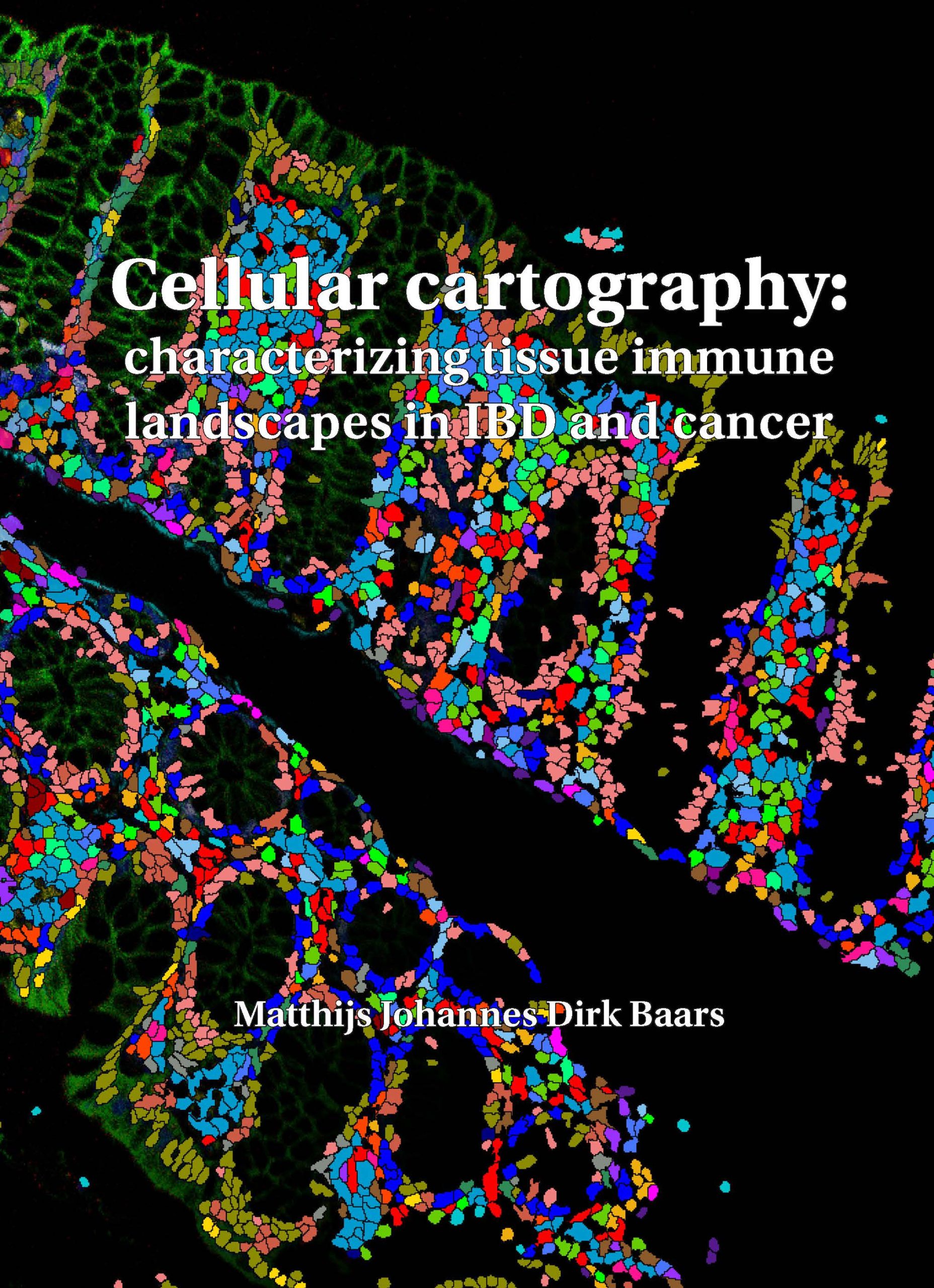
Using an optimized Imaging Mass Cytometry (IMC) workflow, the researchers generated highly detailed maps of immune cells directly within human intestinal tissue. A key methodological advance was the integration of high-resolution fluorescence microscopy to accurately identify individual cells prior to IMC analysis. This approach enabled unbiased and precise single-cell segmentation, even in densely packed tissues such as the intestinal mucosa, and formed the basis for subsequent analyses in disease contexts.
Measuring inflammatory signatures
IMC was applied to intestinal biopsies from patients with long-standing IBD, a group at increased risk of developing colitis-associated cancer. By analyzing inflamed tissue, dysplastic lesions and early tumor stages, the study revealed that the inflammatory immune signature characteristic of IBD persists into early cancerous tissue. Notably, elevated IL-17–associated signaling, a known driver of IBD pathology, was also detected in dysplastic regions. In addition, immune–epithelial interactions were altered during early malignant transformation: cytotoxic T cells were present but positioned further away from the epithelium, suggesting reduced immune surveillance at a critical stage of cancer development. These findings indicate that chronic inflammation not only precedes but actively shapes a tumor-promoting microenvironment.
“Our findings demonstrate that not only the presence but also the spatial organization and activity of immune cells are critical determinants of cancer progression.”
Spatial distribution of cytotoxic T cells
Subsequently, the focus shifted to sporadic colorectal cancers with microsatellite instability (MSI), a subtype characterized by high mutational burden and immune infiltration. Despite abundant immune cells, not all MSI tumors are effectively controlled. By comparing primary tumors from patients who did or did not develop metastases, the researchers found a striking spatial difference in cytotoxic T cell behaviour. Tumors that later metastasized showed cytotoxic T cells remaining at a distance from tumor cells, whereas non-metastasizing tumors displayed dense, active T cell populations in close contact with malignant cells.
Matthijs Baars summarized his findings as follows: “Our findings demonstrate that not only the presence but also the spatial organization and activity of immune cells are critical determinants of cancer progression. This spatial immune profiling approach holds promise as a predictive tool to identify patients at increased risk of tumor progression or metastasis, potentially guiding more personalized surveillance and treatment strategies.”
PhD defense
Matthijs Baars, MSc (1994, Sliedrecht) defended his PhD thesis on January 6, 2026 at Utrecht University. The title of his thesis was “Cellular cartography: characterizing tissue immune landscapes in IBD and cancer.” Supervisor was Prof. Boudewijn Burgering, PhD and co-supervisor was Yvonne Vercoulen, PhD (both Center for Molecular Medicine, UMC Utrecht). Matthijs Baars works data steward & IT admin at the Hubrecht Institute in Utrecht.