Women with dense breast tissue are more likely to develop breast cancer. With the help of AI (artificial intelligence), researchers from UMC Utrecht have now found a factor that may be an additional indicator that cancer is developing within dense breasts: the extent to which the normal glandular tissue lights up on the MRI because of the contrast fluid. This result may ultimately help to more effectively use additional MRI scans to detect cancer in dense breast tissue.
Lees artikel in het Nederlands >
Every two years, Dutch women aged between 50 and 75 are invited to the breast cancer population screening. They can have a mammography made in order to detect breast cancer in an early stage. With about 8 per cent of these women, a mammography is not sufficient because they have so-called ‘dense’ breasts.
This means that their breasts mainly consist of mammary glands and connective tissue instead of fat tissue. As a result, tumours are harder to detect with a mammography. This poses a problem because 50- to 75-year-old women with very dense breasts are three to six times more likely to get breast cancer than the average woman with predominantly fat tissue in her breasts.
Besides increased breast density, there are, of course, many more risk factors involved in breast cancer. These include, amongst other things, obesity, heredity and lifestyle (e.g. smoking, alcohol). A lesser-known risk factor is elevated BPE (background parenchymal enhancement).
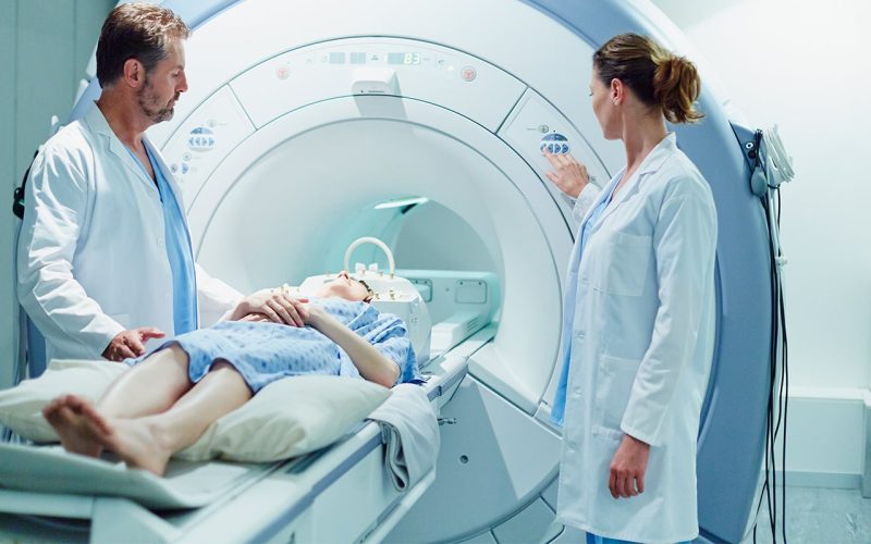
What does this ‘BPE’ mean? During an MRI scan, contrast fluid is injected so that the tumour is more clearly visible compared to healthy tissue. This is because tumours tend to have a better blood flow. But the fluid also enhances ‘normal’ glandular tissue. The BPE factor indicates how actively the glandular tissue reacts to the contrast medium.
For the new UMC Utrecht study, PhD student Hui Wang from the Image Sciences Institute (department of the Image and Oncology Division of UMC Utrecht) developed an AI computer model. With it, the MRI scans of 4,553 women with dense breasts were analysed, taking various additional risk factors into consideration.
Central question of this research: when are women with dense breasts most at risk of developing breast cancer? Conclusion by Wang and her colleagues: when these women’s breasts also show elevated BPE during an MRI, this may be an additional indicator of cancer development. The results of the study were published this week in the leading journal Radiology.
Kenneth Gilhuijs, associate professor at the Image Sciences Institute: “This is the first time a direct link has been established between BPE and cancer development in women with dense breast tissue. There have been more related studies on BPE, but not specifically in women with dense breasts. And this is the first time AI has been used to objectify the results.”
Thanks to the computer model, there was also no need to choose one particular interpretation of elevated BPE. “BPE is a bit of a complicated term because researchers use different definitions for when BPE is elevated. Our computer model can process all interpretations simultaneously,” Gilhuijs explains.
Wang and her colleagues had an impressive amount of MRI scans at their disposal, ready to be used for their AI research. These scans had some years earlier been made during the DENSE trial.
During DENSE, some 5,000 women with dense breast tissue were followed during population screening between 2011 and 2016. These women received an additional MRI scan. The outcomes of this group were compared with the results of the women who only received a mammography (in accordance with the standard population screening). This well-known, elaborate study was coordinated by Carla van Gils, professor of clinical epidemiology of cancer at UMC Utrecht (Julius Centre).
The promising final verdict of the 2020 DENSE study: breast cancer is indeed detected more often and more quickly in women with dense breasts when they receive an additional MRI scan. Furthermore, the use of MRI was also found to be cost-effective.
However, an additional MRI may also have disadvantages. The scan could reveal abnormalities that later turn out to be harmless. Such a false-positive result could cause a lot of stress for women and could lead to unnecessary and costly follow-up examinations and treatments. Capacity problems could also arise: tens of thousands of women would then have to get an MRI in a hospital every year, requiring additional equipment and staff. That is why the Dutch Ministry of Health, Welfare and Sports is having an alternative (contrast mammography) examined first, as advised by the Dutch Health Council.
The new AI study could help to use MRI more efficiently, for instance in population screening. That is, deploying AI to assess MRI scans could save a lot of time and manpower. And if an initial MRI scan shows that a woman has dense breasts but little elevated BPE, it could, for example, be decided that this woman will need less repetitive scans than a woman with both dense breasts and highly elevated BPE. This way, MRI scans could be used in a more targeted and personalised way, which would also reduce the need for staff and equipment.
Gilhuijs: “Our study is a first step towards further personalising the frequency of additional MRI scans in women with dense breasts. This is made possible because, in one glance, we don’t only look at breast density but also at the other risk factors found during the first MRI.”
For cancer care and research, UMC Utrecht is a member of Oncomid. This is a close partnership between hospitals in the centre of the Netherlands. Internationally, the Utrecht region is renowned for its oncological innovations, for instance in the field of image-guided techniques (such as MRI), AI and organoids (‘mini organs’). Within Oncomid, we strive to make all our innovations suitable for future healthcare, which is sustainable, affordable and more manageable for healthcare professionals.
