Functional brain imaging and neurovascular response modeling are two key pillars of ultra-high field (7 Tesla) MRI research. Functional brain imaging uses advanced techniques like laminar fMRI to capture real-time changes in blood flow, volume, and oxygenation with sub-millimeter precision, providing unparalleled insights into neuronal activity. Complementing this, neurovascular response modeling employs computational biophysical simulations to integrate vascular architecture and hemodynamic responses, predicting how these factors influence fMRI signals. Together, these approaches enhance our understanding of brain function across different cortical regions and species, driving forward the field of neuroscience.
For general inquiries, please contact scientific lead Natalia Petridou.
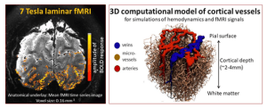
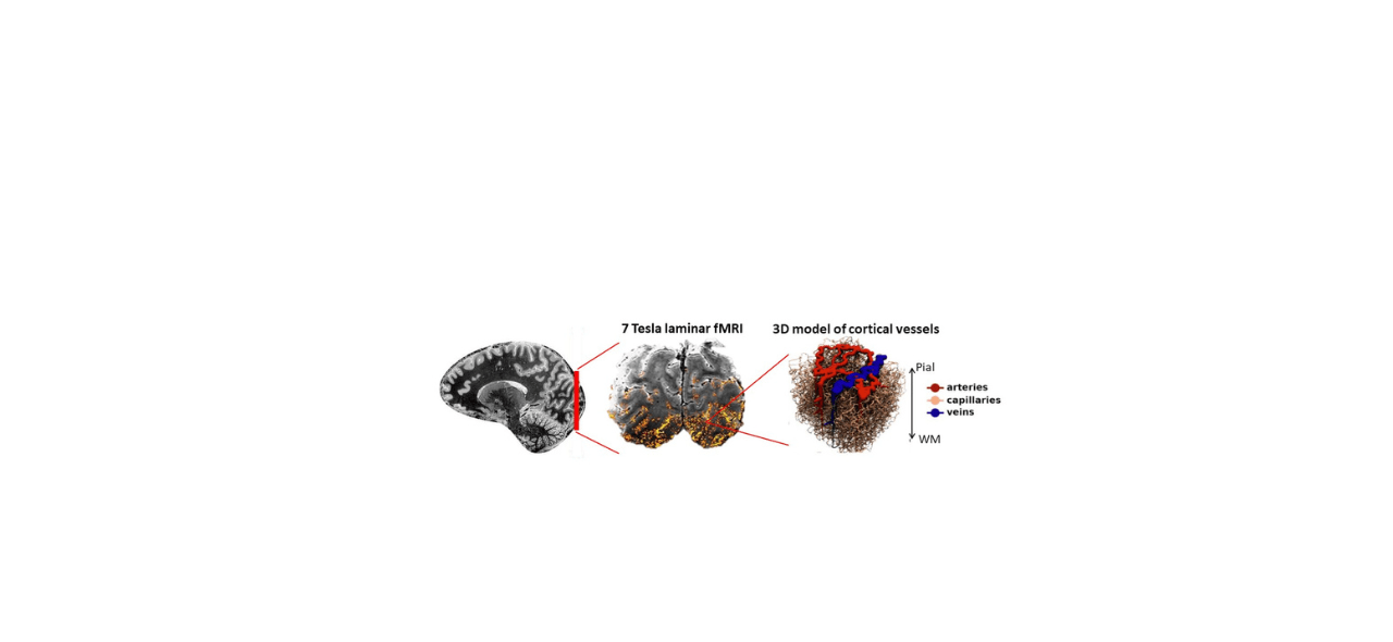
Time series imaging of hemodynamics (blood flow, volume, oxygenation) in response to changes in neuronal activity and metabolism. Contrasts used: Blood-oxygenation-level-dependent (BOLD), cerebral-blood-volume (CBV), and cerebral blood flow (CBF). Imaging techniques: T2*/T2-weighted accelerated echo-planar-imaging (EPI), Vascular space occupancy (VASO), arterial spin labelling (ASL). Laminar fMRI is performed with sub-millimeter spatial resolution.
Computational biophysical modelling that integrates the cortical vascular architecture and hemodynamic response (blood flow, volume, oxygenation), and predicts how these factors determine fMRI measurements. The model creates a synthetic 3D representation of the cortical vasculature based on histological data, and simulates a) hemodynamic parameters (e.g. hematocrit, blood viscosity, flow, volume, oxygenation) and b) biophysical interactions between blood and tissue to characterize physiological contributions to the fMRI signal. The model’s versatility extends to different cortical regions and species, enabling cross-species comparison and study of neurovascular dynamics.