The immune system and organ transplantation: why some organs are accepted and others rejected
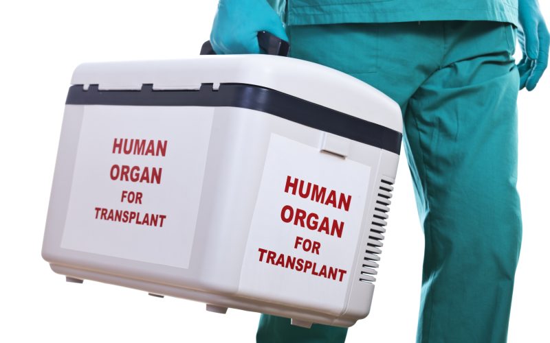
Every year, thousands of patients receive a life-saving organ transplant. A healthy donor kidney, heart or liver can restore organ function and offer patients a second chance at life. But while the operation itself may take only a few hours, the real challenge begins afterwards. Whether the transplanted organ will still function five, ten or even twenty years later depends largely on the recipient’s immune system.
For the immune system, every transplanted organ poses the same fundamental question: does this tissue belong here, or should it be eliminated? For more than half a century, transplant physicians have carefully matched donors and recipients to improve the chances of a successful transplant. This has dramatically improved transplant outcomes and saved countless lives. But even the best-matched organs can still be rejected. Why?
Over the past decade, researchers have realized that not all molecular differences between donor and recipient are equally important. Some patients retain a functioning transplant for decades despite several differences, while others lose a seemingly well-matched organ within a few years. Rather than responding to the number of differences, the immune system reacts to the specific molecular features it can recognize.
Understanding which molecular differences are truly immunogenic is changing the way donor and recipient compatibility is assessed. Rather than relying solely on conventional matching, researchers can now estimate how an individual’s immune system is likely to respond to a transplanted organ.
The challenge of transplantation
Although transplanted organs are healthy tissues that restore organ function, the immune system recognizes them as foreign. Immune cells cannot distinguish between a life-saving donor organ and other forms of foreign tissue.
The immune system consists of two closely connected arms. The innate immune system provides a rapid, first line of defense, while the adaptive immune system mounts a more specific response involving T cells and B cells, two types of specialized white blood cells. Together, these systems shape every stage of transplantation: from the earliest inflammatory response following surgery to the highly specific recognition of donor tissue that can determine whether a transplanted organ is accepted or rejected.
Finding the best donor involves balancing many factors, including blood group compatibility, organ availability, and clinical urgency. Immunological compatibility is one of the most important considerations, and understanding how the immune system recognizes a transplanted organ has become one of the central challenges in transplantation medicine. Even with careful donor selection, however, a match is rarely perfect, and the immune system may still recognize the transplanted organ as foreign.
To prevent rejection, transplant recipients often require lifelong immunosuppressive medication. These drugs suppress the immune response sufficiently to protect the transplanted organ, but they also reduce the body’s ability to fight infections and certain cancers. Modern transplantation therefore depends on a delicate balance: suppressing harmful immune responses while preserving protective immunity.
The body’s molecular passport
Every cell in our body carries an identity card. This identity card consists of Human Leukocyte Antigen (HLA) molecules, proteins displayed on the surface of almost every cell. Their job is surprisingly simple: continuously present small fragments of proteins (peptides) from inside the cell to the immune system. Healthy cells display the body’s own peptides, virus-infected cells display viral peptides, and cancer cells often present abnormal peptides created by mutations. These molecular snapshots are constantly inspected by T cells, that act as the immune system’s surveillance force. By scanning the peptides presented by HLA molecules, T cells determine whether a cell is healthy or poses a threat that should be eliminated. This continuous surveillance allows the immune system to detect infections and cancer at an early stage.
The system is extremely effective, but it comes at a price. The genes encoding HLA molecules are the most diverse in the entire human genome. Thousands of HLA variants have been identified, making it extremely unlikely that two unrelated individuals share exactly the same HLA profile.
From an evolutionary perspective, this diversity is a major advantage. Different HLA molecules present different peptides, meaning that a virus able to evade one person’s immune system may still be recognized by another’s. This genetic diversity has helped protect human populations against infectious diseases throughout evolution.
For transplantation, however, the same diversity creates a formidable challenge. A transplanted kidney does not simply introduce a new organ into the body; it also introduces thousands of unfamiliar HLA molecules. To the recipient’s T cells, these molecules may appear foreign, setting in motion an immune response that, if left unchecked, can ultimately damage or even destroy the transplanted organ.
When “self” becomes “foreign”: how transplant rejection develops
One of the remarkable features of transplantation is how strongly the immune system reacts to donor tissue. In fact, recipient T cells often respond more vigorously to donor HLA molecules than they do to many viruses or bacteria.
The reason lies in how T cells are educated. During their development in the thymus, they learn to tolerate the body’s own HLA molecules, but never someone else’s. As a result, donor HLA molecules can activate large numbers of recipient T cells, triggering a powerful immune response.
Immediately after transplantation, donor antigen-presenting cells activate recipient T cells by displaying intact donor HLA molecules. This process, known as direct allorecognition (from the Greek allos, meaning “other”), drives much of the acute immune response during the first weeks after transplantation. Over time, indirect allorecognition becomes increasingly important. Recipient antigen-presenting cells process donor HLA molecules into peptides, which are then presented to T cells. This pathway plays a central role in long-term immune activation and chronic rejection.
These immune mechanisms help explain why transplant rejection occurs in different forms. Hyperacute rejection develops within minutes to hours after transplantation and is caused by pre-existing antibodies, usually directed against incompatible ABO blood group antigens or donor HLA molecules. Thanks to antibody screening and crossmatching, this form of rejection has become rare. Acute rejection typically develops within weeks or months and is primarily driven by activated T cells, whereas chronic rejection evolves over years as persistent immune activation leads to fibrosis, progressive tissue damage and eventual loss of organ function.
T cells are not acting alone. Once activated, they also provide help to B cells, which produce antibodies against donor HLA molecules. By binding to the blood vessels within the transplanted organ, these antibodies promote inflammation and contribute to progressive tissue damage. Today, both T cell-mediated rejection and antibody-mediated rejection are recognized as major causes of organ injury and are classified according to the internationally adopted Banff Classification, the standard for diagnosing transplant rejection.
Anticipating the immune response
Although T cell-mediated rejection and antibody-mediated rejection are driven by different immune mechanisms, they share a common starting point: the recognition of donor HLA molecules by the recipient’s immune system. Understanding which HLA-derived peptides activate T cells, and subsequently stimulate B cells to produce donor-specific antibodies, has therefore become one of the major challenges in modern transplantation immunology. These discoveries raised an important question: if researchers understand which immune mechanisms drive rejection, can they also predict them before transplantation takes place?
One of the researchers leading this effort is Eric Spierings, associate professor at the Center for Translational Immunology (CTI) at UMC Utrecht and medical immunologist at the Central Diagnostics Laboratory. Together with his team, he combines immunology and computational biology to predict how the recipient’s immune system will respond before transplantation takes place.
“For decades, transplantation has largely been about reacting to rejection. Our goal is to predict who is at risk before transplantation, so we can make better donor choices and ultimately prevent rejection rather than treat it.”
This work led to the development of PIRCHE (Predicted Indirectly Recognizable HLA Epitopes), a computational platform that estimates how likely the recipient’s immune system is to respond to a donor organ.
The clinical impact of this approach is significant. Today, the PIRCHE platform is used by hundreds of transplant centers worldwide. By predicting which donor-derived peptides are most likely to trigger an immune response, it provides information beyond conventional HLA typing and supports donor selection for kidney, liver, heart and lung transplantation.
From reaction to prediction: how AI is changing transplantation
For decades, transplantation medicine has been largely reactive. Patients receive immunosuppressive medication, clinicians monitor organ function, and treatment is adjusted if signs of rejection appear. Today, researchers are trying to turn that approach around. Rather than treating rejection after it has started, they aim to predict which patients are at greatest risk before damage occurs.
This shift is driven by advances in computational immunology. High-resolution HLA sequencing, single-cell technologies and gene-expression analyses generate enormous amounts of data, revealing ever greater detail about how the immune system responds to a transplanted organ. Analizing these complex datasets would be impossible without computational approaches.
Artificial intelligence and machine learning help integrate these different data layers, identifying patterns that would be impossible to detect by conventional methods alone. Instead of replacing clinicians, tools such as PIRCHE support clinical decision-making by providing a more personalized estimate of each patient’s immunological risk.
Ultimately, the goal is simple: moving from reacting to rejection towards predicting and preventing it.
Beyond HLA
Throughout this article, HLA has taken center stage, because it remains the single most important determinant of transplant compatibility. Yet research increasingly shows that HLA is only part of the picture. Whether a transplanted organ is accepted or rejected depends on a complex interplay of immunological factors.
The immune response does not begin only after donor and recipient meet. The transplantation procedure itself also causes tissue damage. When blood flow to the donor organ is temporarily interrupted and later restored, damaged cells release danger signals that activate the innate immune system. This process, known as ischemia-reperfusion injury, triggers inflammation and can amplify the later adaptive immune response.
Genetic differences outside the HLA region also contribute. Minor histocompatibility antigens can trigger T-cell responses even when donor and recipient are perfectly HLA matched, particularly after hematopoietic stem cell transplantation.
Natural killer (NK) cells, part of the body’s innate immune system, and non-HLA antibodies are also recognized as contributors to transplant outcomes. The recipient’s own immune profile, including previous pregnancies, blood transfusions, earlier transplants, and underlying immune status, further influences the risk of rejection.
Together, these findings illustrate that transplant rejection cannot be explained by HLA alone. Rather, it results from the combined effects of donor genetics, recipient immunity, and the biological processes triggered by transplantation itself.
Looking ahead
For more than half a century, HLA molecules have formed the foundation of transplantation immunology. Understanding their role transformed donor matching and made organ transplantation a routine clinical reality.
Today, researchers recognize that transplant compatibility is far more complex than matching HLA alone. The immune response is shaped by a combination of donor and recipient genetics, innate and adaptive immune mechanisms, tissue injury and the individual’s immunological history. Computational models now help integrate these layers of information into a more complete picture of transplant compatibility.
Every transplanted organ still poses the immune system the same fundamental question: does this tissue belong here, or is it foreign? More than half a century after the discovery of HLA, researchers are closer than ever to understanding how the immune system answers that question, and how that knowledge can be used to give every transplant the greatest possible chance of lasting a lifetime.