Imaging in motion: With a €20 million grant from the Dutch Research Council (NWO), the BioMotive consortium, operating at University Medical Center Utrecht and the University of Twente, is developing a new generation of MRI scanners that allow researchers to study the human body during natural movement, such as walking or cycling.
Many diseases are related to posture and movement, from osteoarthritis to cardiovascular diseases and recovery after surgery. Yet, MRI scans are still typically taken while people lie still and flat.
“The BioMotive infrastructure gives us a unique inside view of the body in action. We’ll soon be able to observe how muscles, organs, and metabolism interact during movement, and gain a better understanding of how the body functions in both health and disease.”
Two unique MRI facilities
BioMotive will establish two research facilities:
An open 0.5 Tesla MRI scanner for upright imaging at the University of Twente (to be completed in 2026);
A unique 3 Tesla “walk-in” MRI scanner at UMC Utrecht, enabling imaging during motion (to be completed in 2030).
Both scanners will be equipped with specialized research tools, such as cycling setups and heart monitors, to allow and measure physical exertion. These facilities will enable researchers to study the body in natural postures using MRI, allowing for more precise measurements of how the heart, muscles, and organs work together. The facilities are designed specifically for scientific research, not for direct clinical care.
New insights into heart, muscles, and metabolism
The BioMotive project focuses on three key research areas:
Cardiovascular disease, such as heart failure and hereditary conditions.
The musculoskeletal system, including muscular disorders, sports injuries, and osteoarthritis.
Muscle metabolism and digestion, in the context of metabolic or neuromuscular diseases.
By combining advanced MRI technology with physical modeling, motion analysis, and smart, innovative data processing, researchers will, for the first time, be able to simultaneously map internal forces, blood flow, and muscle tension. “Thanks to BioMotive, we can now observe the body in real-time during realistic movement. That’s truly a breakthrough in biomechanical research,” says Dr. Martijn Froeling (UMC Utrecht).
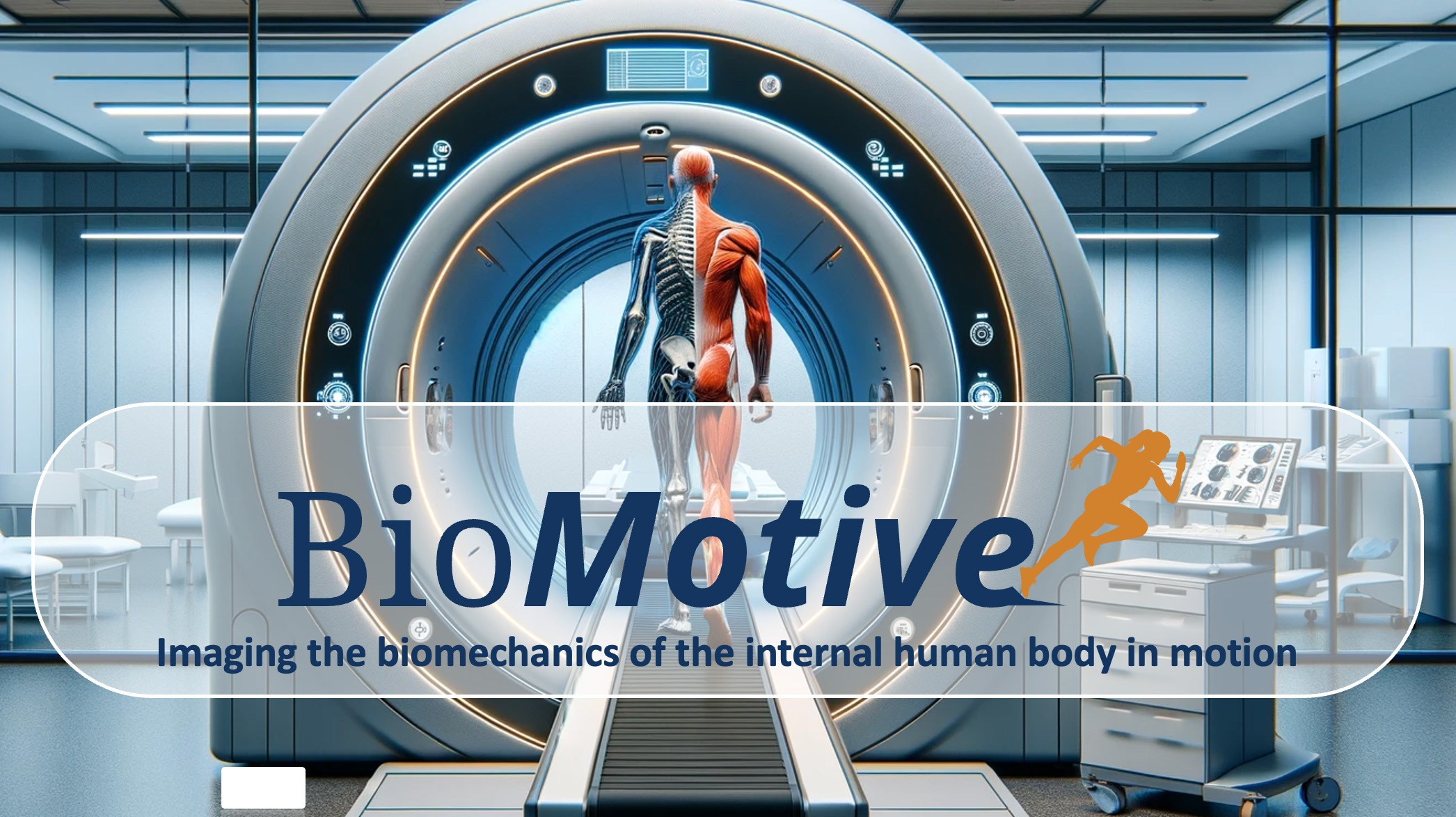
AI illustration of “walk-in” MRI scanner at UMC Utrecht, enabling imaging during motion.
Collaboration without borders
The BioMotive consortium unites leading Dutch knowledge institutions, including UMC Utrecht, University of Twente, Radboudumc, Eindhoven University of Technology, Amsterdam UMC, Maastricht UMC+, UMC Groningen, Wageningen University & Research, Leiden University Medical Center, Erasmus MC, VU Amsterdam, and partners such as Klimmendaal, Kortradio, and the Central Military Hospital.
“This is truly a joint effort,” says Dr. Frank Simonis (University of Twente). “By combining technical MRI expertise with knowledge of advanced biomechanics and cardiology, we are creating something that no single party could have achieved on its own.”
What are the goals of BioMotive?
The goals of the new BioMotive infrastructure are:
To create a platform for broadly applicable, interdisciplinary research.
To gain insights into biomechanical processes and metabolism during natural movement.
To study cardiac function, muscle activity, and metabolism under physical stress.
To combine imaging of motion and metabolism in a single research setup.
To generate new insights for prevention, diagnostics, and treatment.
To support personalized biomechanical models.
Ultimately, to translate these scientific insights into new diagnostic tools, clinical treatments, and guidelines.
About the grant
BioMotive has received €20 million in funding through the Large-Scale Research Infrastructure (LSRI), National Roadmap Consortia 2024 from NWO. This investment will lead to a unique research facility that opens the door to a better understanding of the human body in motion.