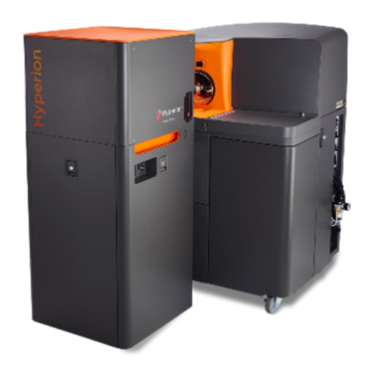
Multiplexed antibody-based single-cell mass spectrometry imaging. The Hyperion™ Imaging System.
The Hyperion™ Imaging System is a high-plex imaging system capable of analyzing 40-plus protein markers at subcellular resolution in frozen or formalin-fixed, paraffin-embedded (FFPE) tissue sections. With the ability to utilize up to 135 channels to detect additional parameters, the Hyperion Imaging System is ideal to meet researcher needs today and well into the future.
The system is based on the Helios™ mass cytometry platform. This high-dimensional imaging platform uses mass-tagged antibodies and employs proven CyTOF® technology.
Mass tagging involves the separation of signals based on the differences in mass, not wavelength, resulting in distinct signals for each marker without the need for compensation associated with fluorometric techniques. The use of metal tags is far more specific and sensitive than tag-free techniques.
The metal tags can be defined within 1 µm spatial resolution in tissue and cell smears, resulting in a unique spatial and parametric definition of the cells in situ. The system enables understanding of protein behavior and interactions to drive biological breakthroughs.
Facilities where you can find this equipment:
| Description | Specification | Description | Specification |
|---|---|---|---|
| channels | 135 | cross-cell contamination | ≤2% at 200 pixels/sec |
| mass range | 75-209 amu | crosstalk pixel to pixel | ≤15% at 200 pixels/sec |
| abundance sensitivity | 0.3% for 159Tb | wet tissue thickness for full ablation | ≤7 µm |
| frequency | 200 pixels/sec | addressable sample size on slide | ≥15 mm × 45 mm |
| detection limit | ≥400 copies per µm2 | optical view of sample | ≥250 µm × 250 µm |
| dynamic range | 4 orders of magnitude | file type | TXT, multipage TIFF, OME-TIFF, MCD |
| scan area | ≥1 mm2/2 hr (@200 Hz) |
