Research approaches in neuro-oncology at the UMC Utrecht Brain Center encompass optimization of anti-tumor treatment, including surgery, radiotherapy, and systemic therapy, alongside investigation into predictors for cognitive symptoms and the development of novel techniques. These efforts involve translational research, cohort studies, neuroimaging, and collaboration with other institutions, aiming to enhance treatment efficacy, patient monitoring, and understanding of tumor biology.
We aim to optimize anti-tumor treatment for brain tumor patients: surgery, radiotherapy and systemic therapy, both traditional chemotherapy and novel drugs. Further fields of interest include tumor-related epilepsy, liquid biopsies for brain tumors including CNS lymphomas and gliomas, and several other clinical research areas. In several cohort studies, we look into the effects of optimal resective neurosurgery, often ‘awake craniotomies’.
In further translational research, we try to unravel the mechanisms behind resistance of gliomas to anti-tumor-treatment. To this end, a new technique called ‘organoids’ is being developed in the laboratory, with the goal to simulate actual tumors. These organoids form a model in which we can study the biological behavior of gliomas. In the future, those organoids can be used to test whether – existing or experimental – treatments are effective to treat a tumor. We also look for the oncobiological mechanisms behind several clinical symptoms, including epilepsy and cognitive symptoms.
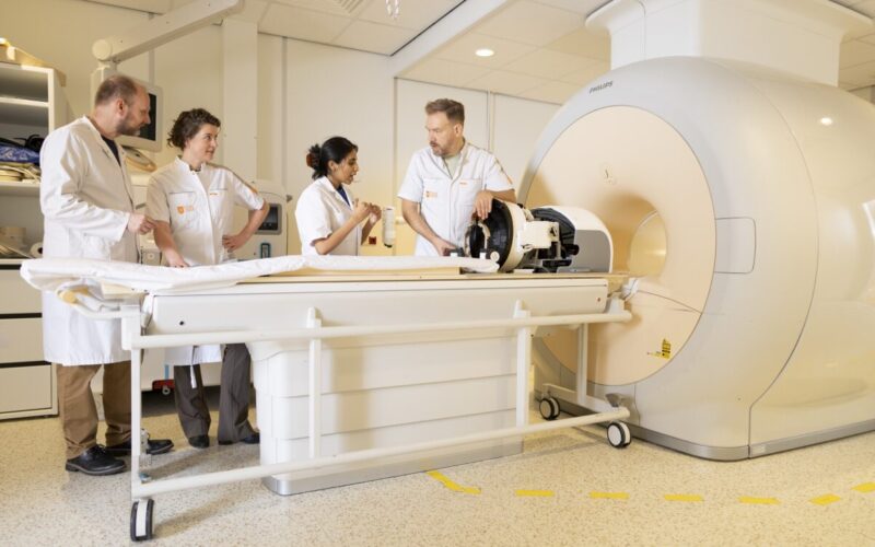
Cognitive symptoms are common in glioma patient. We study the clinical characteristics and predictors for cognitive dysfunction through epidemiological and imaging studies, and correlate these findings to molecular tumor characteristics. To unravel the basis of cognitive symptoms in patients with brain tumors, we run several neuroimaging studies. We aim to correlate tumor and (radiation) treatment characteristics on the one hand, to structural and functional brain imaging on the other. Resting-state fMRI, DTI, high-field (7 Tesla) anatomic MRI, perfusion imaging are some of the techniques used. Ultimately, these data are linked to patients’ cognitive functioning, as measured with detailed neuropsychological testing.
High-frequency focused ultrasound (HiFU) is a promising novel technique to improve blood-brain-barrier passage of drugs. This should lead to a better effect of medication against brain tumors and other brain disease. We collaborate with the Prinses Maxima Center for pediatric oncology to study this technique.
In the follow-up of brain tumor patients, MRI scans are the most-used method of monitoring the tumor. Adequate response assessment turns out to be difficult: when a tumor seems to grow on MRI, this may represent progression of the tumor, but it may also be an effect of treatment (‘pseudoprogression’). We aim to improve radiological follow-up, and to reliably identify and manage pseudoprogression.