Back to Research-topics

Cellular and molecular mechanisms in the injured newborn brain

By understanding the pathophysiological mechanisms of neonatal brain injury, we aim to find novel targets for treatment and ultimately improve neurodevelopmental outcomes in patients.

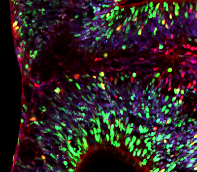
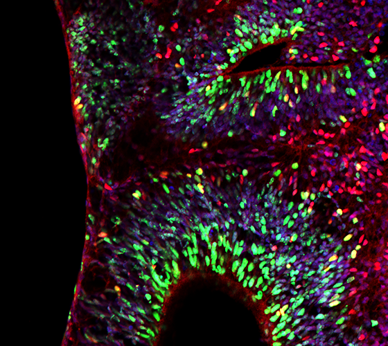
Image right: Cerebral organoid stained for proliferating cells (red, Ki67) and oligodendrocytes (green, Olig2), DAPI is used as nuclear counterstain. Courtesy of Myrna Brandt.

Our research focuses on understanding the pathophysiology of brain injury in the neonatal period, which is a major contributor to mortality and lifelong morbidity. In our translational research group we study how early life events, like hypoxic-ischemic perinatal events, including birth asphyxia and perinatal stroke, or (extreme) preterm birth, lead to injury in the developing brain and how this impacts neurodevelopmental outcome on the long run.

Deciphering Early Brain Injury Mechanisms to Develop Neuroprotective Strategies for Newborns

Our team explores the pathophysiological cellular and molecular mechanisms of early brain injury in in vitro and in vivo models relevant for the target patient. In one of our research lines we try to decipher early damaging cascades after hypoxic-ischemic injury in the term brain. Understanding the intricate interplay between these detrimental cascades can aid in identifying novel targets to develop new strategies to combat neuronal brain injury in the newborn brain. We currently focus on targets within neuronal apoptotic cascades, mitochonadidrial key proteins and neuroinflammatory routes.

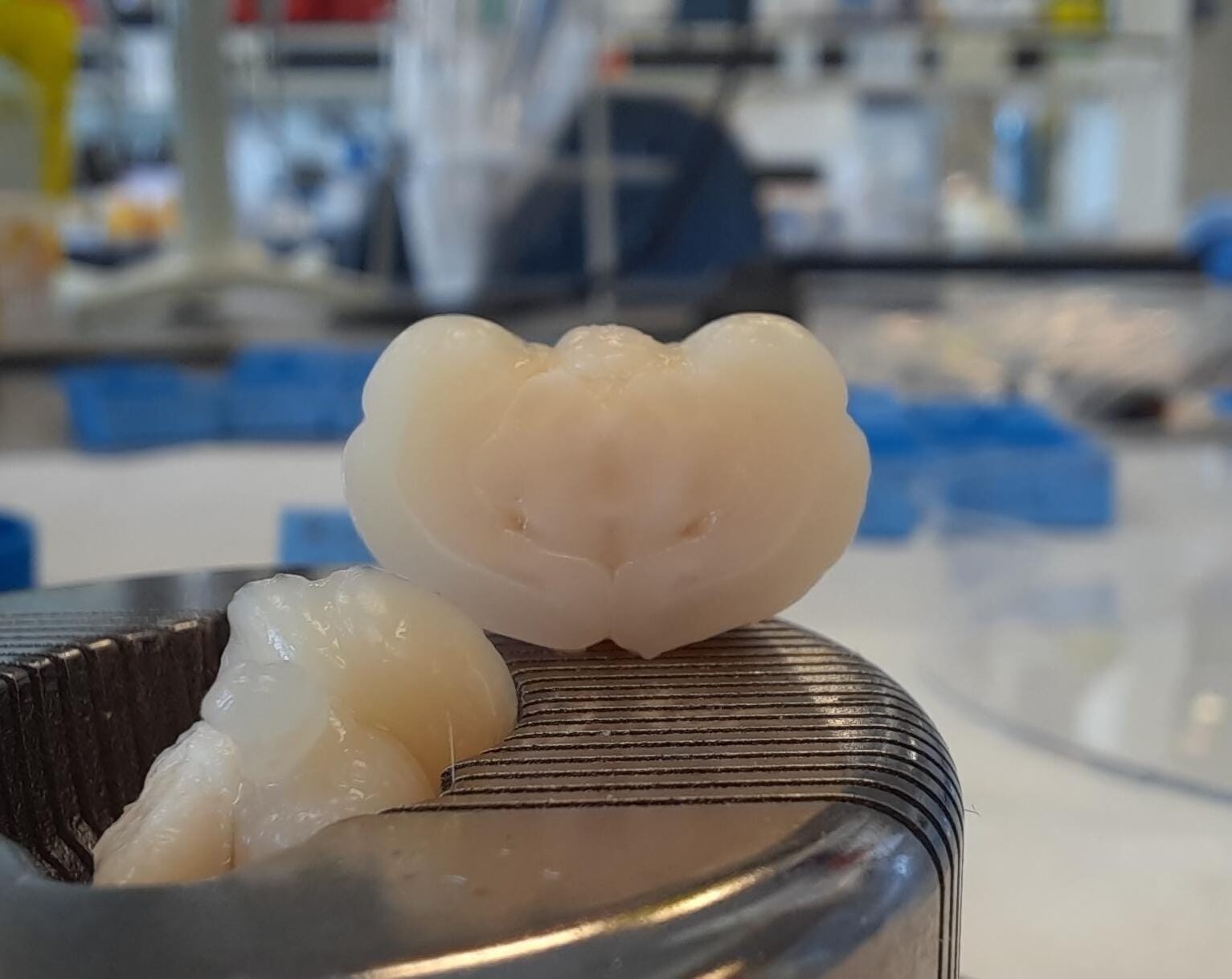
Image: Fixed rat pup brain ready for embedment in paraffin. Courtesy of Judit Alhama Riba & Sebastiaan Corstjens.

Advancing Models and Therapies for Brain Injury following Extreme Preterm Birth

In an additional research line, we focus on developing in vitro and in vivo models for brain injury after extreme preterm birth (i.e. encephalopathy of prematurity (EoP)) including cerebral organoids. We study the underlying pathophysiology of EoP which comprises of hypomyelination of the developing brain, maturational arrest of oligodendrocyte precursors, aberrant interneuron development and neuroinflammation. We currently investigate whether stem cell therapy, growth factor supplementation, human milk oligosaccharide supplementation or immunomodulation would be promising strategies to boost myelination in the preterm brain.


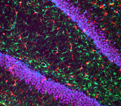
Image left: Brain of a rat exposed to fetal inflammation and postnatal hypoxia as a model for encephalopathy of prematurity, showing active microglia (red, Iba-1) and astrocytes (green, GFAP) in the hippocampus (neuronal nuclei in blue). Courtesy of Chantal Kosmeijer.

Image right: Cerebral organoid stained for proliferating cells (red, Ki67) and oligodendrocytes (green, Olig2), DAPI is used as nuclear counterstain. Courtesy of Myrna Brandt.

  • ZonMw Meer Kennis, Minder Dieren (2023): Timing of fetal exposure to inflammation steers developmental brain injury in the newborn rat.
  • ZonMw Meer Kennis, Minder Dieren (2023): Experimental models of Fetal Growth Restriction: a systematic review on neurological outcome.
  • Valliant (2022): Schade aan de corpora mammilaria tijdens de geboorte: Is het te voorkomen?
  • 3V stimuleringsfonds (2020): Organoïde-model voor hersenschade bij te vroeg geboren kinderen (Myrna Brandt).
  • ZonMw Meer Kennis, Minder Dieren (2018): Witte stof hersenschade in neonatale ratten leidt niet tot verminderde cognitieve vaardigheden op volwassen leeftijd (Erik van Tilborg).
  • Brain Foundation the Netherlands (2016), the Next Step program: Intranasal growth factor treatment: a novel strategy to repair the injured preterm brain.
  • Brain Foundation the Netherlands (2014), fellowship: Mesenchymal stem cell therapy to repair white matter injury in the preterm neonatal brain: boosting oligodendrocyte differentiation and myelination.
  • WKZ Research Funding (2013): A novel strategy to protect the preterm brain against perinatal white matter injury.
Contact

PI’s: Cora Nijboer and Caroline de Theije

Prof. Dr. Cora Nijboer
Personal profile page
LinkedIn page
Follow Cora on X
Email: C.Nijboer@umcutrecht.nl


Dr. Caroline de Theije
Personal profile page
LinkedIn page
Email: C.G.M.deTheije@umcutrecht.nl


Key publications

Ultrasonic vocalization emission is altered following neonatal hypoxic-ischemic brain injury in mice Eva C. Hermans, Caroline G.M. de Theije, Cora H. Nijboer, E.J. Marijke Achterberg Behav Brain Res. 2024; 471:115113
Combined fetal inflammation and postnatal hypoxia causes myelin deficits and autism-like behavior in a rat model of diffuse white matter injury. Van Tilborg E, Achterberg EJM, van Kammen CM, van der Toorn A, Groenendaal F, Dijkhuizen RM, Heijnen CJ, Vanderschuren LJMJ, Benders MNJL, Nijboer CH. Glia 2018; 66:78-93.
Impaired oligodendrocyte maturation in preterm infants: potential therapeutic targets. Van Tilborg E, Heijnen CJ, Benders MJ, van Bel F, Fleiss B, Gressens P, Nijboer CH. Progress Neurobiol. 2016; 136:28-49.
Targeting the p53 pathway to protect the neonatal ischemic brain. Nijboer CH, Heijnen CJ, van der Kooij MA, Zijlstra J, van Velthoven CT, Culmsee C, van Bel F, Hagberg H, Kavelaars A. Ann Neurol. 2011; 70(2):255-64.
Strong neuroprotection by inhibition of NF-kappaB after neonatal hypoxia-ischemia involves apoptotic mechanisms but is independent of cytokines. Nijboer CH, Heijnen CJ, Groenendaal F, May MJ, van Bel F, Kavelaars A. Stroke. 2008; 39(7):2129-37.
Back to top